From protection to maturation

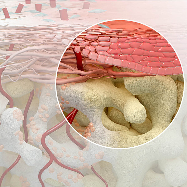
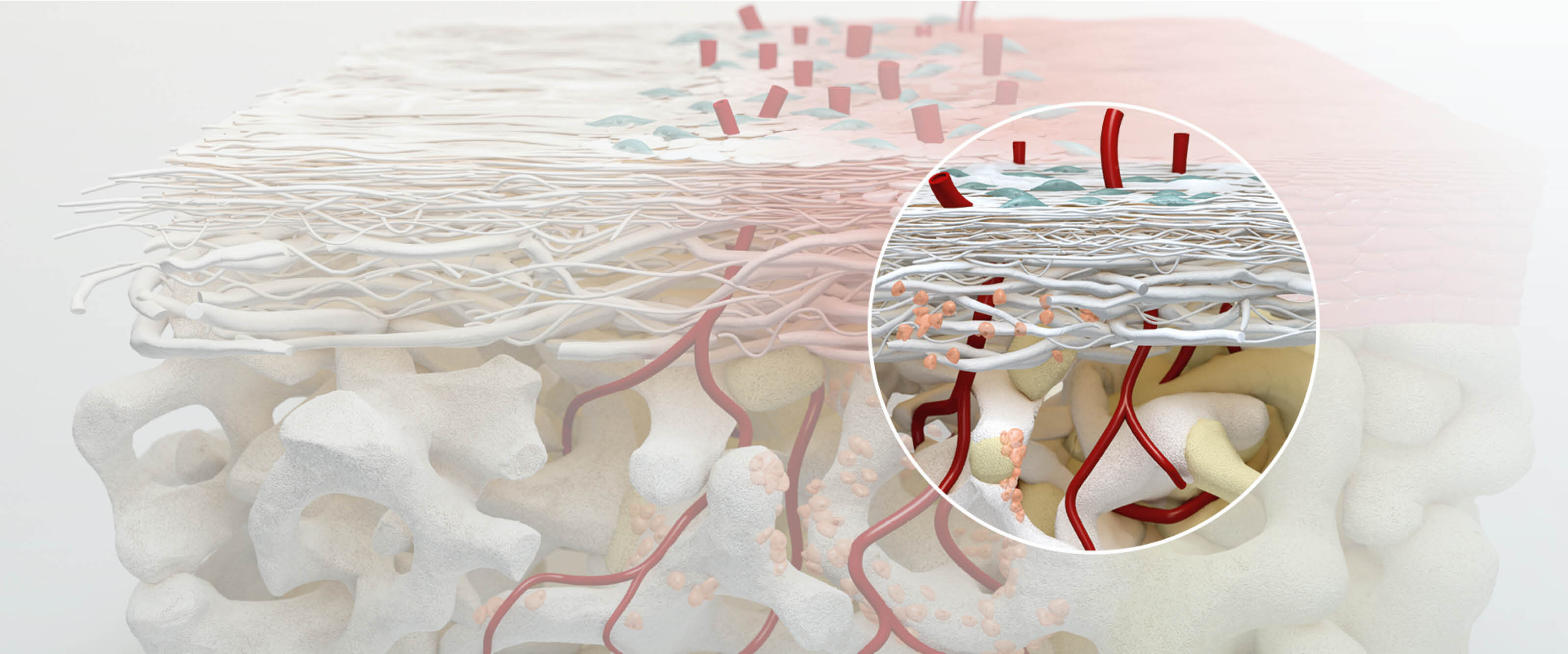
Collagen membranes are used in different treatment protocols including ridge preservation, maxillary sinus lift, or filling bony defects around the dental implant. Vascularization is one of the most important factors in the prognosis of treatment outcomes in the augmented site.1
How long does it take for a collagen membrane to be fully vascularized?
A precise answer to this question requires conducting a clinical study with repeated removal of biopsies for histological analysis. While such practice should be justified for ethical reasons, scientists have tackled the question through animal studies.2-3
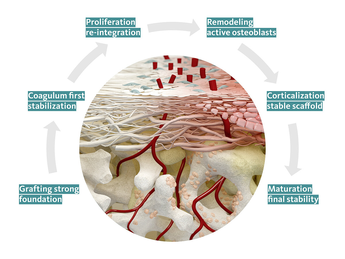
In 2005, Rothamel et al. published the results of a study on the integration of different collagen membranes in a rat model.2 They compared the biodegradation, vascularization, and tissue integration of eight commercially available or experimental collagen membranes. For the Geistlich Bio-Gide®, the histometric analysis showed that the membrane thickness reduced between weeks 2 and 4 (p<0.001), with no significant change for the rest of the observation period (p>0.05). The membrane was completely vascularized at week 2 with the blood vessels reaching the non‐exposed surface of the membrane. It also showed obvious tissue integration at week 2, until the membrane was nearly completely degraded at week 4. While all the other membranes studied had a longer biodegradation time than Geistlich Bio-Gide®, the authors concluded that the “prolonged biodegradation seemed to be associated with decreased tissue integration, vascularization and also foreign body reactions.”2
Later in 2008, Schwarz et al. published the results of a dog study to answer a similar question.3 They placed the implants in the healed ridge in the mandible of 12 beagle dogs, filled the defect with Geistlich Bio-Oss®, and covered it with six different membranes or left it uncovered. Healing was uneventful in all dogs with no complications during the observation period. However, the time of ingrowth of blood vessels through the thickness and thus the resorption time varied for each membrane. For Geistlich Bio-Gide®, the newly formed blood vessels had homogeneously penetrated through the histological specimens at week 1. At week 4, the thickness of the membrane was significantly decreased (p<0.05), followed by the increased mean bone fill value measured at week 6 (p<0.05). Finally, at week 12, Geistlich Bio-Gide® was almost resorbed, and a significant increase in the mean bone formation was observed (p<0.01).
Does vascularization promote regeneration?
An established and mature vascular network assists and accelerates the regenerative processes by bringing the blood supply and the required nutritional elements and growth factors to the augmented site.1
What do the clinical studies say?
Geistlich Bio-Gide® has been used by clinicians for at least two decades:4-5more than 80 clinical studies report successful treatment outcomes with Geistlich Bio-Gide®. As Prof. Daniel Buser says, “The Geistlich Bio-Gide® membrane has proven itself to be reliable and is characterized by easy clinical handling and a low complication rate for 20 years.”
References:
- Saghiri MA, et al.: Med Oral Patol Oral Cir Bucal 2016;21(4):e526-e537. (Review)
- Rothamel D, et al. : Clin Oral Implants Res 2005;16(3):369-78. (Pre-clinical study)
- Schwarz F, et al.: Clin Oral Implants Res 2008;19(4):402-15. (Pre-clinical study)
- Camelo M, et al.: Int J Periodontics Restorative Dent 1998;18(4):321-31. (Clinical study)
- Jung RE, et al.: Clin Oral Implants Res 2013;24(10):1065-73. (Clinical study)
About the author

Manager Medical Communications
Geistlich Pharma AG