![](/fileadmin/content/RegenerationExpert/Artikel/12_Various/Pispero_Clinical_case_2023/Clinical_Case_Regenfast_Header_2400x1000_Pispero.jpg)
Boosting oral surgery with REGENFAST®
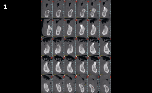
Regenerative treatment of a site after osteonecrosis of the jaw
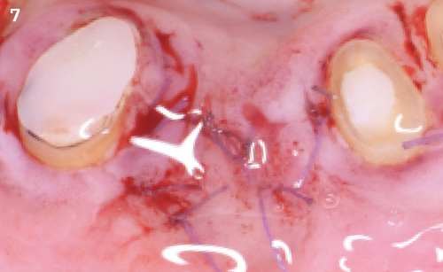
"I used REGENFAST® for the healing of the surgical site. Polynucleotides and hyaluronic acid seem to speed up soft-tissue healing. This is essential for improved bone healing in patients at risk of medication-related osteonecrosis of the jaw."
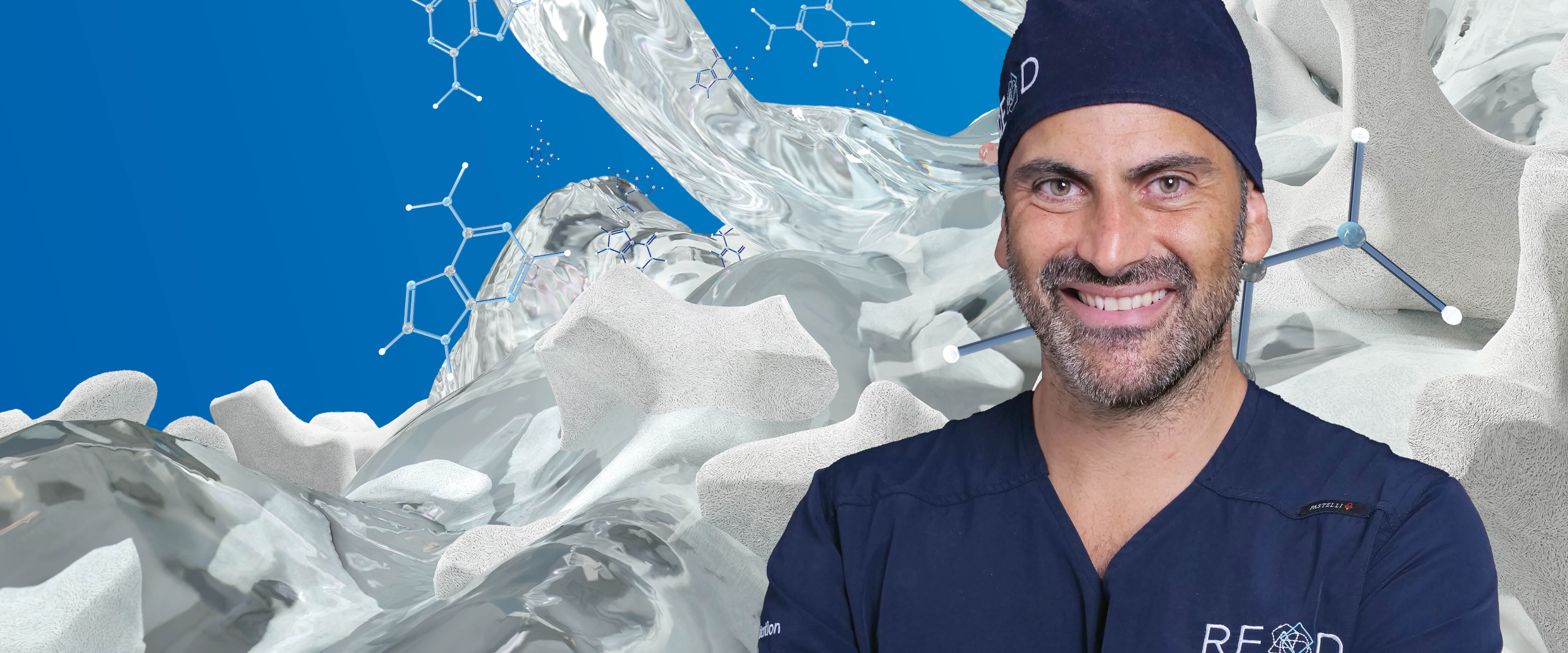
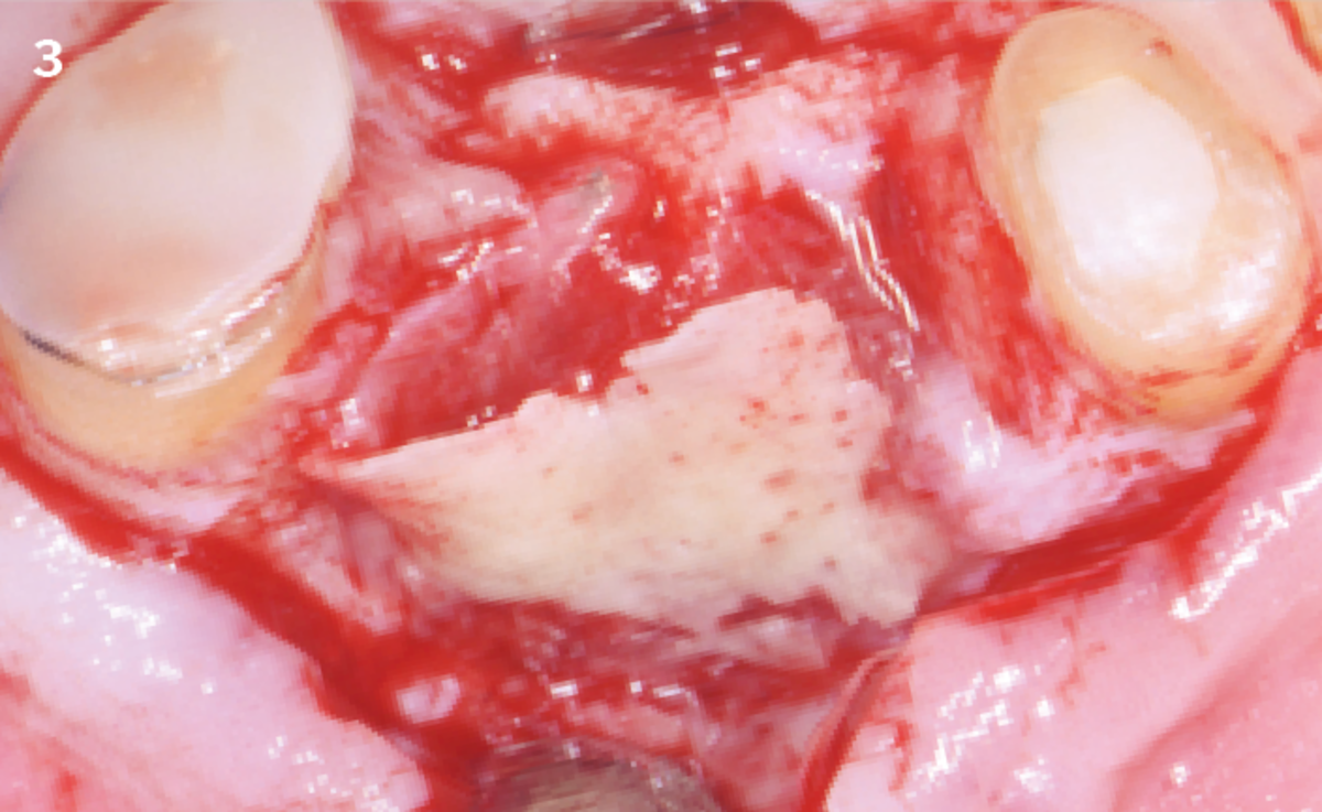
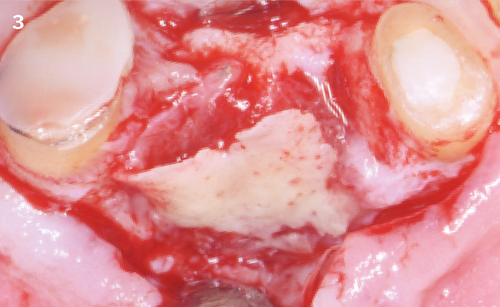
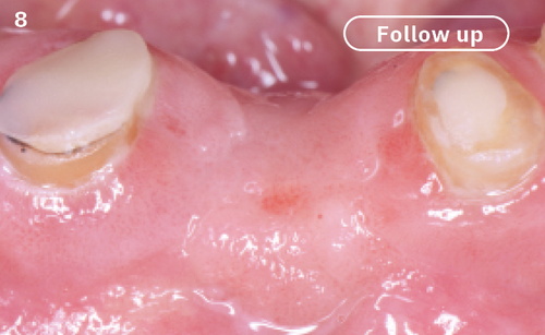
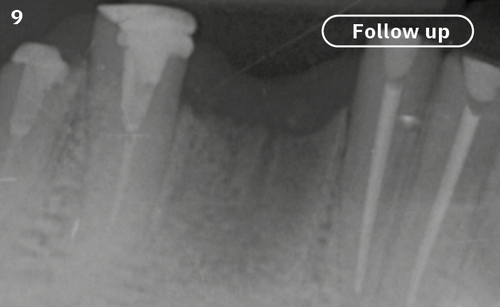
The Situation
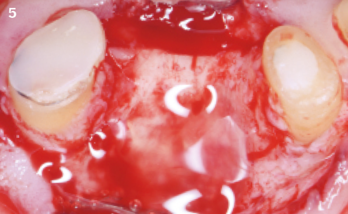
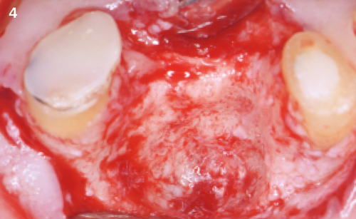
Following the extraction of tooth 42, a drug-induced osteonecrosis was diagnosed and treated. One week after the treatment, the clinical picture had improved and the symptoms were significantly reduced.
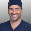
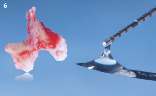
Approach & Result
![1 | Cone beam sections show a fragment of
necrotic bone.](/fileadmin/_processed_/f/5/csm_Pispero_01_ef95440c8c.png)
![2 | Fistula in relation to the edentulous crest through which the bone crest can be probed.](/fileadmin/_processed_/6/0/csm_Pispero_02_d4cb9715fd.png)
![3 | Intraoperative image of the necrotic bone fragment, corresponding to the vestibular cortex of the element previously extracted.](/fileadmin/_processed_/5/5/csm_Pispero_03_8bf29b2c90.png)
![4 | The removal of the fragment is simple because of complete sequestration.](/fileadmin/_processed_/8/4/csm_Pispero_04_47f769f117.png)
![5 | The clinically healthy bone tissue is curetted and a generous layer of REGENFAST® is applied
before wound closure by primary intention.](/fileadmin/_processed_/c/1/csm_Pispero_05_46a7ef0001.png)
![6 | The viscosity of the product allows it to remain in the intervention site. Contact with saliva would cause rapid dispersion in the oral cavity.](/fileadmin/_processed_/3/1/csm_Pispero_06_84c8c30eec.png)
![7 | After the suture, the excess part of the product is applied on the suture line.](/fileadmin/_processed_/9/4/csm_Pispero_07_bd4c391e3c.png)
![8 | Perfectly healthy tissues with no signs of pathology three months after the surgery.](/fileadmin/_processed_/5/a/csm_Pispero_08_43b169be5b.png)
![9 | The intraoral radiographic image confirms the bone healing of the treated site.](/fileadmin/_processed_/3/4/csm_Pispero_09_fe8d1d3956.png)
![1 | Cone beam sections show a fragment of
necrotic bone.](/fileadmin/_processed_/f/5/csm_Pispero_01_32de291cb5.png)
![2 | Fistula in relation to the edentulous crest through which the bone crest can be probed.](/fileadmin/_processed_/6/0/csm_Pispero_02_39287c6b3d.png)
![3 | Intraoperative image of the necrotic bone fragment, corresponding to the vestibular cortex of the element previously extracted.](/fileadmin/_processed_/5/5/csm_Pispero_03_3f2558dac3.png)
![4 | The removal of the fragment is simple because of complete sequestration.](/fileadmin/_processed_/8/4/csm_Pispero_04_c9d2651d0f.png)
![5 | The clinically healthy bone tissue is curetted and a generous layer of REGENFAST® is applied
before wound closure by primary intention.](/fileadmin/_processed_/c/1/csm_Pispero_05_8517dbc227.png)
![6 | The viscosity of the product allows it to remain in the intervention site. Contact with saliva would cause rapid dispersion in the oral cavity.](/fileadmin/_processed_/3/1/csm_Pispero_06_b7e849af6e.png)
![7 | After the suture, the excess part of the product is applied on the suture line.](/fileadmin/_processed_/9/4/csm_Pispero_07_968141a4fc.png)
![8 | Perfectly healthy tissues with no signs of pathology three months after the surgery.](/fileadmin/_processed_/5/a/csm_Pispero_08_8590495a74.png)
![9 | The intraoral radiographic image confirms the bone healing of the treated site.](/fileadmin/_processed_/3/4/csm_Pispero_09_510ab2fc6e.png)
Download the Case