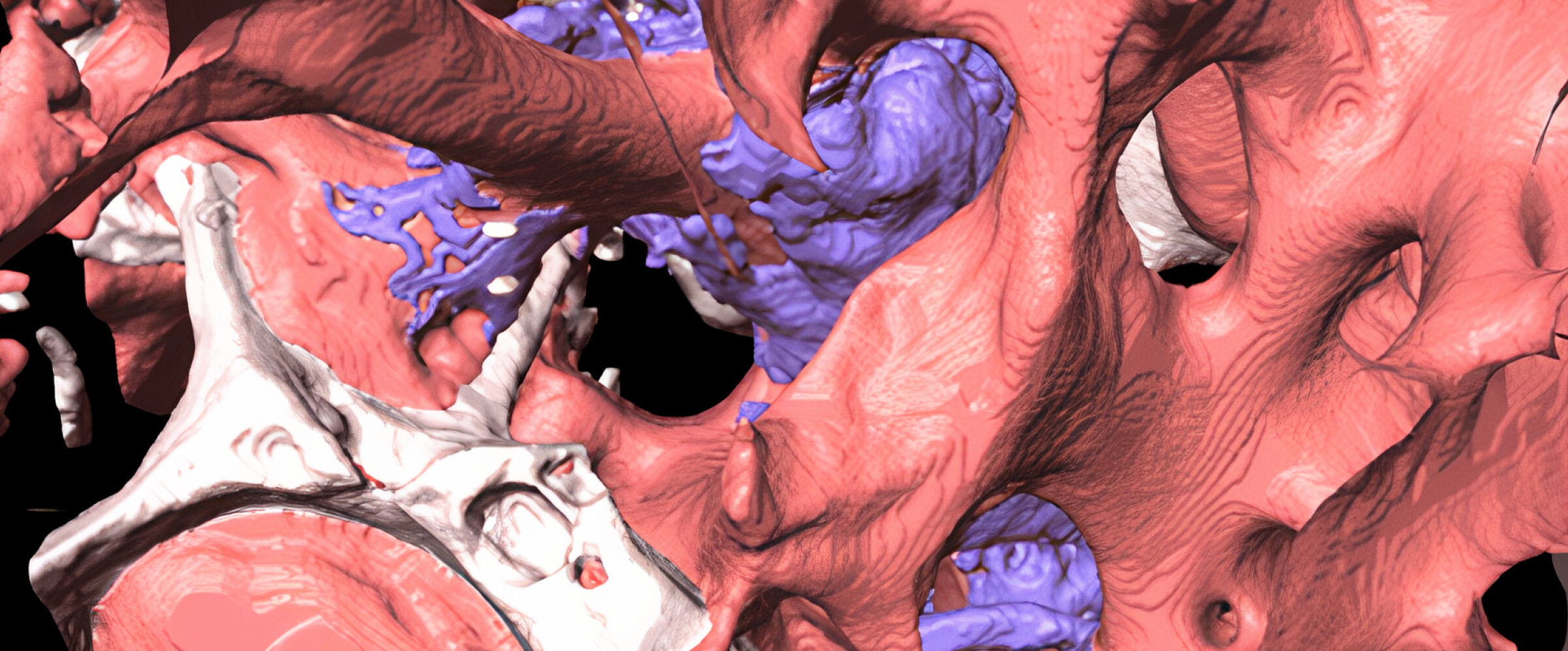
Navigation inside the 3D pore structure of regenerated bone

Synchrotron Radiation micro-computed tomography (SR-μCT) is based on high flux X-ray in a particle accelerator. The acquired images have a high resolution and allow simultaneous visualization of the specimen’s 3D microstructure and quantitative analysis of the segments, along with their densities. With SR-μCT the intrinsic limitations of traditional histomorphometric microcopy, e.g., the 2D nature of images or specimen defects during sectioning, are surmounted. It is also possible to study the bone regeneration dynamic, including the ratio and distribution of new and substitute bone and their interfaces at different times, which is not possible using conventional µCT or historical histomorphometry.
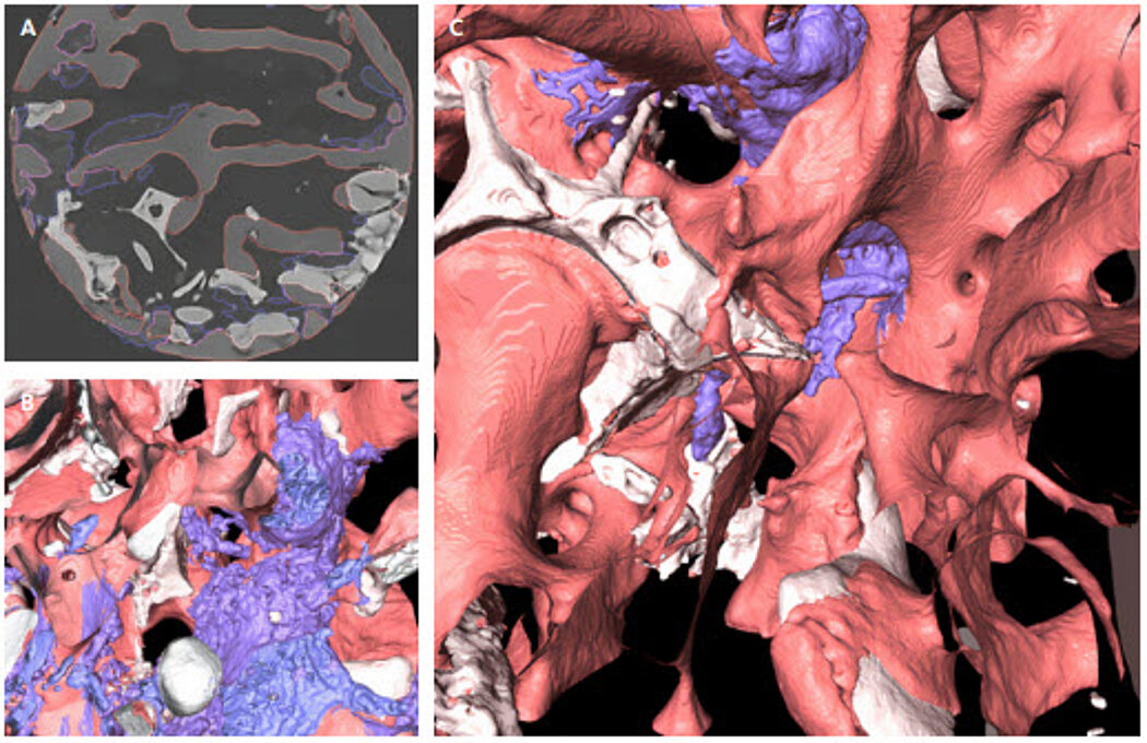
In our study, sinus floor augmentation was performed using the lateral approach and Geistlich Bio-Oss®.¹ After 6 months of uneventful healing, the bone biopsy specimens were collected during implant site preparation and cut into a cylindrical shape for tomography image acquisition (3 mm long and 2 mm diameter). After reconstructing the 3D images from the tomography slices (Fig. 1A), the true structure of regenerated bone was revealed (Fig. 1B, C). Quantitative analysis indicated the volume fractions of new bone, Geistlich Bio-Oss® and woven bone were 29.44%, 13.39%, 13.29%, respectively.
About the author

Department of Periodontology, School of Dentistry,
Kyungpook National University, South Korea