Advanced Ridge Augmentation with 3D Printed Titanium Scaffold

The Situation
A 34-year-old female was referred for rehabilitation of her partially edentulous maxillary dentition; a situation which she has endured since her early teenage years. She has been wearing a removable partial denture ever since failure of her fixed dental bridgework. The patient was keen on an implant solution to manage her situation, originally requesting an all on-four implant rehabilitation of her maxillary arch. She was medically healthy, a non-smoker, and taking oral contraceptive medication. Clinical examination revealed extensive horizontal and vertical ridge deficiencies with a knife-edge ridge.
The Approach
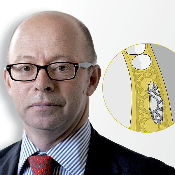
The goal was to re-establish sufficient ridge volume for dental implant rehabilitation. Advanced postextraction ridge resorption necessitated both horizontal and vertical augmentation. Due to the large extent of the defect and need to ensure long-term volume stability, a slowly resorbing bovine bone augmentation material (Geistlich Bio-Oss®) was combined with autologous bone to establish the foundation for bone regeneration. The mixture was stabilised with an Yxoss CBR® titanium scaffold.
A resorbable collagen barrier membrane (Geistlich Bio-Gide®) was draped over the scaffold to ensure separation of the osseous and soft tissues during the early healing phase. The underlying regenerating bone was well-supported and protected by Yxoss CBR® and Geistlich Bio-Gide®. The intaglio surface of the removable dental prosthesis was relieved such that minimal pressure would be applied to the healing soft tissues.
The Outcome
The patient presented with an extensive bone defect making ridge reconstruction particularly challenging. Use of the volume-stabilising Geistlich Bio-Oss® shaped by the innovative Yxoss CBR® 3D titanium scaffold enabled reconstruction of the ridge to the correct anatomical form. Despite a minor dehiscence of the flap during the healing phase, which sometimes occurs, the ridge augmentation was successfully achieved in a controlled and efficient manner.
Keys to Success
- Stabilization of 3D titanium scaffold and membrane
- Flap design and tension-free closure
- Use of deproteinized bovine bone mineral with low substitution rate and autologous bone (50:50 ratio)
- Importance of keratinized tissue for soft tissue stability
- Relief of provisional prosthesis to reduce pressure on the underlying healing tissues
About the author

BDS Hons (SYD), Grad Dip Clin Dent (Oral Implants) (SYD), M Clin Dent (Pros) (LON), D Clin Dent (Pros) (SYD)
Dr. Christopher Ho is a Specialist Prosthodontist with a Bachelor of Dental Surgery with First Class Honours at the University of Sydney. He completed postgraduate studies in the Graduate Diploma in Clinical Dentistry in Oral Implants, Masters of Clinical Dentistry in Prosthodontics with Distinction from the University of London and Doctorate in Clinical Dentistry in Prosthodontics from University of Sydney. He is Editor of the Wiley textbook “Practical Procedures in Aesthetic Dentistry” and “Practical Procedures in Implant Dentistry.”